
More Information
Submitted: October 01, 2026 | Accepted: October 09, 2026 | Published: October 12, 2026
Citation: Osunwoke EA, Chidera OH, Okon M. Ergonomic Survey of Foot Dimensions and Morphological Patterns in the Ikwerre Population. J Forensic Sci Res. 2026; 10(2): 154-160. Available from:
https://dx.doi.org/10.29328/journal.jfsr.1001125
DOI: 10.29328/journal.jfsr.1001125
Copyright license: © 2026 Osunwoke EA, et al. This is an open access article distributed under the Creative Commons Attribution License, which permits unrestricted use, distribution, and reproduction in any medium, provided the original work is properly cited.
Keywords: Bloodstain pattern analysis; Rough surface; Surface topography; Environmental factors; Crime scene reconstruction
Abbreviatiions: FL: Foot Length; FW: Foot Width; LFL: Left Foot Length; LFW: Left Foot Width; RFL: Right Foot Length; RFW: Right Foot Width; SD: Standard Deviation; SE: Standard Error; SPSS: Statistical Package for the Social Sciences; NS: Not Significant

Ergonomic Survey of Foot Dimensions and Morphological Patterns in the Ikwerre Population
Emeka Anthony Osunwoke1 , Owhor Harmony Chidera1 and Michael Okon2*
1Department of Human Anatomy, Faculty of Basic Medical Sciences, College of Health Sciences, University of Port Harcourt, Rivers State, Nigeria
2Department of Human Anatomy, Faculty of Basic Medical and Allied Sciences, College of Health Sciences, Nigerian British University, Asa, Abia State, Nigeria
*Corresponding author: Michael Okon, Department of Human Anatomy, Faculty of Basic Medical and Allied Sciences, College of Health Sciences, Nigerian British University, Asa, Abia State, Nigeria, Email: [email protected]
Background: Foot morphology varies across populations and is influenced by genetic, environmental, and cultural factors. Understanding the dominant foot shape and differences in foot parameters within this population is important for clinical assessment, footwear design, and anthropological documentation. This study aimed to determine the dominant foot shape among the Ikwerre population and assess sex differences and bilateral asymmetry in foot length and width.
Methods: A cross-sectional study was conducted among 200 participants (100 males and 100 females) aged 18–40 years. Foot scans were obtained using a digital scanner, while foot length and width were measured using a digital vernier caliper. Data were analyzed using SPSS version 16.0, and comparisons were made using t-tests and chi-square tests, with significance set at p<0.05.
Results: Males exhibited significantly greater foot length and width compared to females (p < 0.05). A statistically significant asymmetry was observed between the right and left feet in both measurements. The Egyptian foot shape was the most prevalent in both sexes, followed by the Greek foot type. Foot shape distribution did not differ significantly between males and females (p > 0.05).
Conclusion: The Ikwerre population predominantly exhibits the Egyptian foot shape, with notable sexual dimorphism and mild bilateral asymmetry in foot dimensions. These findings hold relevance for clinical foot care, orthotic and footwear design, and contribute valuable anthropometric data on an underrepresented Nigerian ethnic group.
Foot anthropometry has become increasingly important across clinical biomechanics, orthopedics, podiatric medicine, and ergonomic footwear design as population-level variation is recognized to influence musculoskeletal outcomes. Modern anthropometric practice has progressed from simple linear measurements to three-dimensional scanning and geometric morphometric techniques, enabling more accurate capture of foot shape and form [1]. Foot morphology, shaped by genetic heritage, habitual activity, lifestyle, and environmental exposure, plays a central role in gait mechanics and plantar pressure distribution and is associated with conditions such as hallux valgus, plantar fasciitis, and bone-stress injuries [2-4]. Because foot dimensions and shape profiles vary across populations, locally derived anthropometric data are essential to inform orthotic prescriptions and the design of culturally and anatomically appropriate footwear [1,3]. In this context, documenting foot morphology among the Ikwerre ethnic group will improve clinical assessment, guide ergonomic shoe design, and contribute valuable regional anthropometric data.
While numerous studies have investigated foot morphology among global and African populations, there remains a paucity of research specifically focused on smaller indigenous communities such as the Ikwerre people of Rivers State. Existing literature on foot morphology in Nigeria has predominantly centered on larger ethnic groups such as the Igbo, Yoruba, and Hausa, leaving a gap in the scientific understanding of foot structure, with limited attention given to smaller indigenous communities like the Ikwerre community [5,6]. Given the cultural diversity and ecological differences among Nigerian ethnic groups, localized anthropometric data are essential for accurate clinical evaluations, footwear production, and ergonomic interventions. Therefore, a focused study on the Ikwerre population is necessary to address this gap and provide insights into their unique anatomical characteristics.
The Ikwerre people are an indigenous ethnic group located in the Niger Delta region of Nigeria, primarily inhabiting several Local Government Areas in Rivers State, including Port Harcourt, Obio-Akpor, Emohua, and Ikwerre. They are recognized among the major indigenous nationalities in the region, and they have rich cultural heritage and traditional practices that may influence their foot morphology and foot health. Foot anthropometric studies among Nigerians have demonstrated variation in foot dimensions and shape across different populations and ethnic groups [7,8]. In addition, research on habitual activity patterns and footwear use has shown that minimal or flexible footwear can influence foot strength, structure, and weight-bearing mechanics, further contributing to population-specific morphological differences [9]. Given that the Ikwerre people engage in traditional occupations such as fishing and farming and may adopt minimal or flexible footwear habits, it is plausible that their foot morphology might differ, which this present study seeks to explore. These lifestyle-related factors strengthen the need for a population-specific assessment of foot morphology among the Ikwerre.
Studies show that foot morphology differs between ethnic groups, influenced by genetic, cultural, and environmental factors [10]. Research among Indigenous Arctic populations and other geographically distinct groups indicates climate-related and footwear-related adaptations in forefoot and arch form [11]. In contrast, studies of habitually barefoot East African populations demonstrate longer and narrower foot shapes, associated with long-distance walking and distinctive gait patterns [12,13]. Furthermore, foot morphology and foot problems are influenced by footwear choices, physical activity, and occupation [14]. In addition, a study found that habitual high-heel use has been shown to alter the neuromechanics of walking and is associated with increased risk of musculoskeletal foot problems [15]. These findings highlight the dynamic relationship between lifestyle behaviours and foot anatomy.
In addition, foot morphology can also be related to various medical conditions, such as diabetes, inflammatory arthritis, and neurological disorders, which increase the risk of foot ulcers, deformity, and chronic pain [16,17]. Foot problems are common among people with inflammatory arthritis and often include structural deformities and persistent pain [18].
Several Nigerian anthropometric studies further support the existence of population-specific patterns in foot structure. Musa, et al. [19] examined foot dimensions among the Ga'anda people and found significant sex-based variations, with males showing consistently higher values for foot length, breadth, and height. Similarly, Bhattacharjee, et al. [20] reported substantial differences in foot dimensions between Bangalee and Chakma females, emphasizing the role of ethnicity in determining foot shape and size. These findings align with broader anthropometric trends, demonstrating that foot morphology cannot be generalized across populations.
Studies exploring the relationship between foot dimensions and stature also reinforce the influence of population differences. Oghenemavwe and Egwede (2022) [21] established that foot length is a strong predictor of stature among the Uturu indigenes of Abia State, with sex-specific regression equations highlighting the importance of locally derived anthropometric models. In the Hausa population, Alabi, et al. [22] demonstrated that toe lengths could reliably aid in sex discrimination and contribute to stature estimation models, though with varying degrees of accuracy. Among children, Bafor, et al. [23] found significant correlations between foot length and height in Nigerian schoolchildren, further emphasizing the functional relevance of foot dimensions in forensic and clinical applications.
Other studies have advanced foot morphology research through more sophisticated approaches. Hong, et al. [24] utilized three-dimensional foot scanning to analyze extensive foot shape variables, girth, width, height, and angles, among over 2,000 Chinese young adults. Their results demonstrated significant sex-based differences in foot shape and type, underscoring the utility of advanced measurement techniques in capturing the complexity of foot structure. These findings highlight the evolving methodological landscape in foot anthropometry and provide a comparative foundation for studies using simpler but reliable measurement tools such as calipers and foot scanners.
Foot morphology also has substantial implications for health. Variations in foot structure may contribute to conditions such as arthritis, diabetes-related complications, and neuropathic changes. Armstrong, et al. [17] reported that individuals with diabetes are at heightened risk of foot ulcers due to altered foot loading and peripheral neuropathy, while Boulton, (2008) [16] observed that arthritis and chronic inflammatory conditions can lead to deformities and persistent foot pain. These health-related findings emphasize the need for accurate foot assessments in clinical practice, especially within populations with limited healthcare access.
Therefore, this study aims to address the gap in knowledge by conducting a comprehensive investigation into the dominant foot shape among the Ikwerre people. By employing a combination of anthropometric measurements, data analysis techniques, and demographic surveys, this research seeks to characterize the foot morphology of the Ikwerre population and explore any associations between foot shape, cultural practices, and lifestyle factors. The findings of this study will contribute to the existing literature on foot morphology and provide valuable insights for healthcare professionals, footwear designers, and researchers seeking to address foot health issues and improve the quality of life for individuals within the Ikwerre community. During the study, we hoped to determine the dominant foot shape, explore any correlations between foot shape, cultural practices, or lifestyle factors, and contribute to existing anthropometric data on foot morphology among individuals in the Ikwerre population of Rivers State, Nigeria.
Study design
This study employed a cross-sectional research design to assess foot morphology among a selected sample of the Ikwerre population.
Study population and sample size
A total of 200 participants were recruited for the study, comprising 100 males and 100 females between the ages of 18 and 40 years. The minimum sample size was determined using the Taro Yamane formula [25]:
n = N/((1+N*e^2))
where;
n = sample size
N = population size
e = margin of error (0.08, in this study). The reason why the margin of error was picked at 0.08 was due to the homogeneity of the population.
The estimated population of Ikwerre in Rivers State, Nigeria is 2,000,500 respectively (City Population, accessed 8th Oct. 2026; 11:13 am)
Therefore, N = 2,000,500, e = 0.08
n = 2,000,500/1+2,000,500 (0.08)2
= 156.24
Therefore, approximately 200 subjects.
Sampling method
A purposive sampling method was employed to select individuals of Ikwerre origin from Rivers State.
The Inclusion Criteria for this study were males and females between the ages of 18 to 40 years that are of Ikwerre origin in Rivers State (by paternal and maternal association to the second generation) and eligible to participate in the study, while exclusion criteria were those with obvious foot deformities, history of foot injury and fracture, and surgical procedures affecting the feet.
Anthropometric measurement
For the foot anthropometric parameters, measurements were taken to assess various aspects of foot morphology among the Ikwerres. The following parameters were measured:
Foot length: This was measured using a digital vernier caliper. It is the distance from the most posterior point of the heel to the most anterior point of the longest toe (great or second toe).
Foot width: The widest point of the foot was measured using a digital vernier caliper. It was taken as the distance between the most prominent point on the medial aspect of the head of the first metatarsal (Figure 1) and the most prominent point on the lateral aspect of the head of the fifth metatarsal.
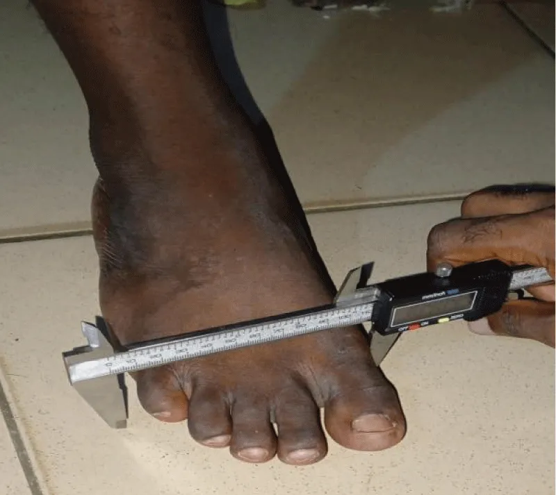
Figure 1: Measurement of foot width.
Overall foot shape classification
Characterized based on visual inspection of foot scans or digital models, considering factors such as arch type, toe alignment, and overall symmetry. The different foot shapes are the Egyptian, the Greek, and the square (also called Roman) foot, as described by Vesin, (2021) [26]
For this study, Egyptian and Greek foot types were predominantly observed and recorded (Figure 2 & Figure 3).
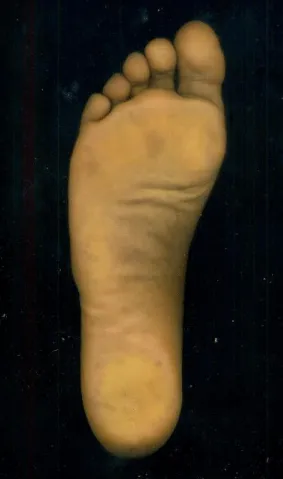
Figure 2: Egyptian foot shape in the studied population.
Figure 3: Greek foot shape in the studied population.
Data collection procedure
Each participant was asked to sit in an upright position on a chair to reduce the effect of weight-bearing on the foot. Each participant's foot was gently placed on a digital HP scanner, then the foot was captured, and their respective shapes were recorded (Figure 4). The length and width of each subject foot were measured afterwards using the digital vernier caliper. All measurements and image acquisitions were performed by a single operator to ensure standardization and reduce intra-observer variability.
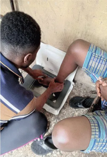
Figure 4: Foot Scanning Procedure.
Limitations of the study
- Limited time for data collection
- Intermittent power supply during scanning procedures
- Possible measurement errors and bias
- Cultural and socioeconomic differences among participants e.g occupation, living conditions and health
Data analysis
Data were analyzed statistically using Statistical Package for Social Sciences (SPSS) version 16.0. The Mean, standard deviation (SD) and standard error (SE) were recorded. An independent samples t-test was used to compare foot length and foot width between both sexes, a paired samples t-test was used to compare the right and left foot of the same participant, and the chi-square test was used to compare the distribution of foot shape between the sexes. Significance was set at p < 0.05.
Ethical consideration
Ethical clearance was gotten from the University of Port Harcourt research ethic committee with the approval number: UPH/R&D/REC/EXEC/221.
A total of 200 participants were included in the study, consisting of 100 males and 100 females, representing 50% each of the total study population (Figure 5). The results of this study are presented in the tables and charts below.
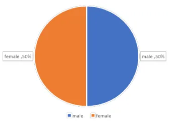
Figure 5: Sex distribution of the study population, with an equal representation of males and females, each comprising 50% of the total sample..
Table 1 summarizes the descriptive statistics of the measured foot parameters.
| Table 1: Descriptive Statistics of Foot Parameters. RFW = Right foot width; RFL = Right foot length; LFW = Left foot width; LFL = Left foot length; | ||||||
| Parameters | N | Min | Max | Mean | SEM | SD |
| RFW (mm) | 200 | 70.69 | 114.32 | 90.93 | .79 | 11.28 |
| RFL (cm) | 200 | 18.70 | 27.50 | 23.75 | .14 | 1.94 |
| LFW (mm) | 200 | 70.23 | 113.42 | 90.36 | .78 | 11.04 |
| LFL (cm) | 200 | 18.70 | 29.20 | 23.59 | .13 | 1.85 |
The mean ± standard deviation (SD) for Right Foot Width (RFW) was 90.93 ± 11.28 mm, Right Foot Length (RFL) was 23.75 ± 1.94 cm, Left Foot Width (LFW) was 90.36 ± 11.04 mm, and Left Foot Length (LFL) was 23.59 ± 1.85 cm.
Sex differences in foot parameters
Sexual dimorphism was observed in the foot measurements. Males had significantly greater foot width and foot length when compared to females (p < 0.05). The results of the comparison are presented in Table 2.
| Table 2: Sex Differences in Foot Parameters. RFW = Right foot width; RFL = Right foot length; LFW = Left foot width; LFL = Left foot length; S = significant (p < 0.05). | |||||
| Parameter | Male (Mean ± SD) | Female (Mean ± SD) | t-test | p - value | Inference |
| RFW (mm) | 98.94±9.65 | 82.92±4.93 | 14.24 | <0.001 | S |
| RFL (cm) | 25.32±1.22 | 22.18±1.08 | 19.23 | <0.001 | S |
| LFW (mm) | 98.29±9.76 | 82.42±4.75 | 14.61 | <0.001 | S |
| LFL (cm) | 25.04±1.22 | 22.12±1.06 | 17.98 | <0.001 | S |
The mean ± standard deviation (SD) for Right Foot Width (RFW) showed a significant difference (M = 98.94 ± 9.65, F = 82.92 ± 4.93, t = 14.24, p < 0.001), Right Foot Length (RFL), Left Foot Length (LFW) and Length Foot Length (LFL) were also significant. Right Foot Length (RFL) (M = 25.32 ± 1.22, F = 22.18 ± 1.08, t = 19.23, p < 0.001). Left Foot Width (LFW) (M = 98.29 ± 9.76, F = 82.42 ± 4.75, t = 14.61, p < 0.001), and Left Foot Length (LFL) (M = 25.04 ± 1.22, F =22.12 ± 1.06, t = 17.98, p < 0.001).
Asymmetry between left and right foot
Table 3 illustrates the asymmetrical differences of the right and left foot in both length and width, and the mean value of FW was (R = 90.93 ± 11.28, L= 90.36 ± 11.04, t = 4.87, p < 0.001) and FL was (R = 23.75 ± 1.94, L= 23.59 ± 1.85, t = 6.27, p < 0.001). The table showed that FW and FL were statistically significant with asymmetry.
| Table 3: Asymmetry Differences Between Right and Left Foot. FL = Foot length; FW = Foot width; S = significant (p < 0.05). | |||||
| Parameter | Right (Mean ± SD) | Left (Mean ± SD) | T-test | p- value | Inference |
| FW (mm) | 90.93±11.28 | 90.36±11.04 | 4.87 | <0.001 | S |
| FL (cm) | 23.75±1.94 | 23.59±1.85 | 6.27 | <0.001 | S |
Prevalence of foot shape
Table 4 showed the prevalence of foot type in the right foot, and it showed that Egyptian foot was predominant foot shape followed by Greek foot shape in males while in female, Egyptian foot was also predominant, and it was accompanied by Greek foot shape. The prevalence of foot shape in the right foot between sexes were not significant (p > 0.05).
| Table 4: Prevalence of foot shape in the right foot. Pearson chi-square test, df = 1; χ² = 2.41; NS = non-significant (p > 0.05) | |||||
| Foot shape | Female n (%) | Male n (%) | Total (%) | p - value | Inference |
| Egyptian foot | 92 (92.0%) | 85 (85.0%) | 177 (88.5%) | 0.121 | NS |
| Greek foot | 8 (8.0%) | 15 (15.0%) | 23 (11.5%) | ||
Table 5 showed the prevalence of foot type in the left foot, and it showed that Egyptian foot was predominant foot shape followed by Greek foot shape in males while in female Egyptian foot was also predominant and it was accompanied by Greek foot shape. The prevalence of foot shape in the left foot was not significant (p > 0.05).
| Table 5: Prevalence of Foot Shape in the Left Foot. Pearson chi-square test, df = 1; χ² = 2.00; NS = non-significant (p > 0.05) | |||||
| Foot shape | Female n (%) | Male n (%) | Total (%) | p - value | Inference |
| Egyptian foot | 93 (93.0%) | 87 (87.0%) | 180 (90.0%) | 0.157 | NS |
| Greek foot | 7 (7.0%) | 13 (13.0%) | 20 (10.0%) | ||
This study was carried out to determine sex differences, asymmetry in foot parameters, and the prevalence of foot shapes amongst the Ikwerre population in Rivers State, Nigeria. The findings from the descriptive and inferential statistics revealed several important patterns related to foot morphology that are consistent with the existing literature on foot dimensions by [27] and contribute new insights to the Nigerian and African foot morphology database. These results also reflect the first known dataset describing foot shape distribution among the Ikwerre people.
Sex differences in foot dimensions
Significant differences between males and females in foot dimensions were observed in this research. The males exhibited greater foot width and foot length compared to females across all measured parameters. The right foot width (RFW) and left foot width (LFW) for males were significantly higher than for females. The mean RFW for males was 98.94 mm compared to 82.92mm for females, which was statistically significant (p < 0.001). Similar trends were observed in right foot length (RFL) and left foot length (LFL). These findings are supported by the work of Nieves, et al. [28], who reported that males tend to have larger foot dimensions across various populations due to differences in bone density and muscle mass.
These differences can be attributed to biological factors, such as the generally larger skeletal structure of males compared to females, which has been widely documented in other anatomical studies [29], and agrees with the findings of Musa, et al. [19] among the Ga’anda population, Bhattacharjee, et al. [20] among Bangalee and Chakma ethnic groups, and Hong, et al. [24] in a Chinese cohort, all of whom reported significantly greater male foot dimensions. Similar observations were made by Alabi, et al. [22] among the Hausa, where males had significantly higher mean toe dimensions than females.
These outcomes are further supported by the recently published study conducted in Ijebu-Ode, Southwest Nigeria, which reported larger foot parameters among males compared to females and emphasized the role of biological and skeletal variation in determining foot size [30]. By reinforcing this trend, the present study aligns with broader anthropometric evidence, suggesting that sexual dimorphism in foot morphology is a globally consistent feature influenced by bone density, muscle mass and overall body habitus.
Foot asymmetry in the ikwerre population
Significant asymmetry was observed between the rights and left feet in both foot width and foot length, with the right foot generally larger than the left. This slight yet significant asymmetry is consistent with the findings by Sforza [31,32], who attributed that foot dominance and asymmetry to habitual activity patterns. The right foot, often dominant in physical activity, is exposed to more force and strain, leading to slight increases in foot dimensions over time [22], also reported subtle side differences in toe lengths among the Hausa, supporting the idea that repeated force transmission on the dominant limb may influence foot morphology over time.
The Ijebu-Ode foot morphology study also documented slight but notable asymmetry in foot dimensions, which was attributed to gait habits, occupational activities, and footwear exposure [30]. The similarity between the Ijebu-Ode findings and the current Ikwerre data highlights a recurring pattern of right-sided dominance in Nigerian populations. Clinically, this reinforces the importance of considering bilateral foot differences in orthotic fabrication, shoe sizing, and biomechanical assessments to reduce the risk of discomfort or injury.
Prevalence of foot shape among the ikwerre people
Regarding foot shape, the Egyptian foot, characterized by a longer first toe, was the most prevalent foot type amongst the study participants, accounting for 88.5% of right feet and 90.0% of left feet. The Greek foot type, in which the second toe is longer, was less common.
This distribution pattern aligns closely with findings from the Ijebu-Ode population, where the Egyptian foot was also the overwhelmingly prevalent morphology, while the Greek foot type appeared at much lower frequencies [30]. The convergence between the two studies suggests that the Egyptian foot may be characteristic of certain Southwestern and South-South Nigerian populations.
There was no significant difference in the distribution of foot shapes between males and females, as the chi-square test yielded non-significant p-values (p > 0.05). These findings suggest that foot shape is likely influenced more by genetic or hereditary factors than by sex. Although data on foot shape in African populations remain scarce, the findings of this study will contribute meaningfully to the anthropometric record on foot shape research. Overall, the results contribute to a better understanding of foot morphology, showing that while there are clear sex-based differences in foot size; foot shape does not vary significantly between sexes. The dominance of the Egyptian foot type observed here provides a foundation for further research and has practical relevance for ergonomic footwear production tailored to the Ikwerre population.
Implications, objectives, and contribution to knowledge
The objectives of this study were to determine sex differences, examine asymmetry, and identify dominant foot shapes in the Ikwerre population. The findings fulfilled these objectives by demonstrating clear sex-based variations, measurable asymmetry, and the prominence of the Egyptian foot type. The results highlight regional consistencies in Nigerian foot morphology. Notably, the fact that both populations show dominance of the Egyptian foot type and significant male and female size differences strengthens the broader anthropometric relevance of these findings [30].
Overall, this study offers valuable insights applicable to clinical practices, orthotic design, ergonomic footwear development, forensic identification, podiatric assessment, and future anthropometric research. By documenting the foot characteristics of a previously understudied population, the research expands the scientific understanding of foot morphology in Nigeria and contributes to a growing body of literature that underscores the importance of ethnic and regional specificity in anthropometry.
Due to the 8% error margin used in this study, our estimates could differ from the population values by up to ± 8% points at 95% confidence, this could be a limitation of the study.
The results of this study clearly demonstrate significant sex differences in foot width and foot length, with males having larger feet than females in both dimensions. These findings underscore the importance of considering sex when designing footwear and ergonomic products. Additionally, the study revealed a small but statistically significant asymmetry between the left and right foot, which could have implications for the design of custom footwear and orthotic devices. The high prevalence of the Egyptian foot shape further reinforces the need for shoe designs that accommodate this foot type, especially in populations where it is dominant.
The study also confirms that foot shape distribution is not significantly influenced by sex, suggesting that genetic and possibly environmental factors play a larger role in determining foot shape. These findings contribute to the broader literature on human foot morphology and have important practical applications in clinical, ergonomic, and athletic contexts.
- Allan JJ, Munteanu SE, Bonanno DR, Buldt AK, Choppin S, Bullas A, et al. Methodological and statistical approaches for the assessment of foot shape using three-dimensional foot scanning: a scoping review. J Foot Ankle Res. 2023; 16:24. Available from: https://doi.org/10.1186/s13047-023-00617-z
- Buldt AK, Forghany S, Landorf KB, Murley GS, Levinger P, Menz HB. Centre of pressure characteristics in normal, planus and cavus feet. J Foot Ankle Res. 2018; 11:3. Available from: https://doi.org/10.1186/s13047-018-0245-6
- Mei Q, Kim HK, Xiang L, Shim V, Wang A, Baker JS, et al. Toward improved understanding of foot shape, foot posture, and foot biomechanics during running: a narrative review. Front Physiol. 2022; 13:1062598. Available from: https://doi.org/10.3389/fphys.2022.1062598
- Lavigne A, Chicoine D, Esculier JF, Desmeules F, Fremont P, Dubois B. The role of footwear, foot orthosis, and training-related strategies in the prevention of bone stress injuries: a systematic review and meta-analysis. Int J Exerc Sci. 2023; 16(3):721-743. Available from: https://doi.org/10.70252/znrs2138
- Ekezie J, Onuoha A, Nwaokoro C. Anthropometric evaluation of foot shape dimensions and patterns for ancestral determination of South-East Nigerians. Int J Forensic Med Investig. 2018; 4(1):12-21.
- Ndukwu CC, Okeke CU, Chime T, Chukwu I. Anthropometric estimation of foot parameters for the Igbos of Nigeria. J Bioeng Biomed Sci. 2021; 11(3):1-6.
- Ibeabuchi MN, Nandi ME, Bello A, Nandi MO. Predictive equations and multiplication factors for stature estimation using foot dimensions of an adult Nigerian population. Egypt J Forensic Sci. 2018; 8(1):58. Available from: https://doi.org/10.1186/s41935-018-0094-2
- Ikpa TF, Egwu AO, Ochulor OC, Nwafor AO. Discriminant function models for determination of sex using anthropometric foot dimensions in an adult Nigerian population. Int J Anat Res. 2019; 7(3.3):6904-6910.
- Curtis R, Mendez-Villanueva A, Bishop C. Daily activity in minimal footwear increases foot strength: implications for foot structure and function. Sci Rep. 2021; 11:1-12.
- Hoey C, Wang A, Raymond RJ, Ulagenthian A, Okholm Kryger K. Foot morphological variations between different ethnicities and sex: a systematic review. Footwear Sci.. 2022;
- Gurney JK, Kuch C, Rosenbaum D, Kersting UG. The Maori foot exhibits differences in plantar loading and midfoot morphology to the Caucasian foot. Gait Posture. 2012; 36(1):157-159. Available from: https://doi.org/10.1016/j.gaitpost.2012.01.013
- D'Aout K, Pataky TC, De Clercq D, Aerts P. The effects of habitual footwear use: foot shape and function in native barefoot walkers. Footwear Sci. 2009; 1(2):81-94.
- Hollander K, van der Zwaard BC, de Villiers JC, Braumann KM, Venter R, Zech A. Growing up (habitually) barefoot influences the development of foot and arch morphology in children and adolescents. Sci Rep. 2017; 7:1-10. Available from: https://doi.org/10.1038/s41598-017-07868-4
- McRitchie M, Branthwaite H, Chockalingam N. Footwear choices for painful feet: an observational study exploring footwear and foot problems in women. J Foot Ankle Res. 2018; 11:23. Available from: https://doi.org/10.1186/s13047-018-0265-2
- Cronin NJ, Barrett RS, Carty CP. Long-term use of high-heeled shoes alters the neuromechanics of human walking. J Appl Physiol. 2012; 112(6):1054-1058. Available from: https://doi.org/10.1152/japplphysiol.01402.2011
- Boulton AJ. The diabetic foot. In: LeRoith D, Vinik AI (eds) Controversies in treating diabetes. Contemporary endocrinology. Humana Press, Totowa. 2008;
- Armstrong DG, Tan TW, Boulton AJM, Bus SA. Diabetic foot ulcers: a review. JAMA. 2023; 330(1):62-75. Available from: https://doi.org/10.1001/jama.2023.10578
- Simonsen MB, Horslev-Petersen K, Coster MC, Jensen C, Bremander A. Foot and ankle problems in patients with rheumatoid arthritis in 2019: still an important issue. ACR Open Rheumatol. 2021; 3(6):396-402. Available from: https://doi.org/10.1002/acr2.11258
- Musa SC, Jude P, Martha OA, Nathan LD. Comparison of the foot height, length, breadth and foot types between males and females Ga'anda people, Adamawa, Nigeria. J Dent Med Sci. 2015; 14(8):89-93.
- Bhattacharjee S, Ashrafuzzaman M, Chakraborty S. Assessment of foot shape in adult females of Bangalee and Chakma ethnic groups. Chatt Maa Shi Hosp Med Coll J. 2020; 19(1):59-62. Available from: https://doi.org/10.3329/cmoshmcj.v19i1.48806
- Oghenemavwe LE, Egwede OB. Estimation of living stature from foot dimensions in Uturu indigenes of Abia State, Nigeria. Eur J Med Health Sci. 2022; 4(1):67-71.
- Alabi AS, Didia BC, Oladipo GS, Aigbogun EO. Estimation of stature from toe lengths: an evaluation of the Hausa population in Nigeria using linear modeling. J Acad Anat Int. 2017; 3(1):36-43.
- Bafor A, Chibuzom CN, Mbanuzuru AV. Correlation between foot length and height in a cohort of 1,677 Nigerian school children. J Niger Acad Med. 2023; 2(1):60-64.
- Hong Y, Wang L, Xu DQ, Li JX. Gender differences in foot shape: a study of Chinese young adults. Sports Biomech. 2011; 10(2):85-97. Available from: https://doi.org/10.1080/14763141.2011.569567
- Yamane T. Statistics: an introductory analysis, 2nd edn. Harper and Row, New York. 1967;
- Vesin P. The different foot shapes and how to fit them. Podexpert. 2021; https://www.podexpert.com/en/blog-pathology-type-feet-pxl-151_159.html. Accessed 10 September 2026.
- Sundip CH, Kadlimatti HS. Correlation of foot dimensions with body weight: a study in young population of central India. Transl Res Anat. 2019; 16:100043.
- Nieves JW, Formica C, Ruffing J, Zion M, Garrett P, Lindsay R, et al. Males have larger skeletal size and bone mass than females, despite comparable body size. J Bone Miner Res. 2005; 20(3):529-535. Available from: https://doi.org/10.1359/jbmr.041005
- Stankovic K, Booth BG, Danckaers F, Burg F, Vermaelen P, Duerinck S, et al. Three-dimensional quantitative analysis of healthy foot shape: a proof of concept study. J Foot Ankle Res. 2018; 11(1):8.
- Osunwoke EA, Ishiola BP, Okon M. Prevalence of Greek and Egyptian foot morphology in Southwest Nigeria using Ijebu-Ode as a case study. Asian J Med Princ Clin Pract. 2025; 8(2):811-819.
- Sforza C, Michielon G, Fragnito N, Ferrario VF. Foot asymmetry in healthy adults: elliptic Fourier analysis of standardized footprints. J Orthop Res. 2088; 16(6):758-765. Available from: https://doi.org/10.1002/jor.1100160619
- Polk JD, Stumpf RM, Rosengren KS. Limb dominance, foot orientation and functional asymmetry during walking gait. Gait Posture. 2017; 52:140-146. Available from: https://doi.org/10.1016/j.gaitpost.2016.11.028