
More Information
Submitted: October 01, 2024 | Approved: October 07, 2025 | Published: October 08, 2025
How to cite this article: Akrep A. Macroscopic Changes Observed after Burning in Incised Wounds on Bones caused by Sharp and Penetrating Instruments. J Forensic Sci Res. 2025; 9(2): 200-204. Available from:
https://dx.doi.org/10.29328/journal.jfsr.1001103
DOI: 10.29328/journal.jfsr.1001103
Copyright license: © 2025 Akrep A. This is an open access article distributed under the Creative Commons Attribution License, which permits unrestricted use, distribution, and reproduction in any medium, provided the original work is properly cited.
Keywords: Anthropology; Bone; Thermal; Fire; Forensic medicine

Macroscopic Changes Observed after Burning in Incised Wounds on Bones caused by Sharp and Penetrating Instruments
Aytunc Akrep*
Department of Medical Sciences, Istanbul University-Cerrahpaşa, Institute of Forensic Medicine and Forensic Sciences, Istanbul, Turkey
*Address for Correspondence: Aytunc Akrep, PhD Student, Department of Medical Sciences, Istanbul University-Cerrahpaşa, Institute of Forensic Medicine and Forensic Sciences, Istanbul, Turkey, Email: [email protected]
Background: Stab and incised wounds are frequently encountered in forensic practice. Burned skeletal remains often complicate the evaluation of such injuries, making it important to understand how fire alters cut marks on bones. This study aimed to investigate the macroscopic changes of sharp-force trauma on bones after exposure to high temperatures.
Methods: Three bovine humeri were incised with a 32 cm non-serrated kitchen knife. The length, width, direction, and interrelationships of each incision were measured with a forensic scale, photographed, and recorded. The bones were then burned in open-air conditions using oak wood, without accelerants, at 460 °C –570 °C for 60 minutes. Following combustion, the incisions were remeasured, and pre- and post-burning characteristics were compared statistically.
Results: Despite advanced thermal destruction, most incisions remained macroscopically visible. Heat exposure caused significant increases in incision length and width (p < 0.05). Linear fissures radiating from the cuts were observed, and the cut margins appeared sharper and drier after burning. In one specimen, thermal fractures disrupted the evaluation of a distal incision.
Conclusion: Sharp-force trauma marks on bones are not completely obscured by fire and may even become more pronounced. However, heat-related expansion of cut dimensions can lead to overestimation of blade size. These findings emphasize the need for caution in interpreting burned skeletal remains and highlight the importance of further research with larger sample sizes to improve forensic accuracy.
Every year, thousands of people are injured or lose their lives as a result of violent acts. Knives, which are widely used in daily life in places such as homes and workplaces for various purposes, are more easily accessible than firearms because they are not subject to specific restrictions at the time of purchase. Due to their constant availability, these instruments frequently appear in forensic medicine practice in cases involving crimes against bodily integrity.
According to the Violence Map of Turkey prepared by the Umut Foundation in 2022, sharp instruments were used in 616 (15.5%) of firearm-related violent incidents that resulted in homicide, while 3,368 (84.5%) of these homicides were committed with firearms [1]. With the parallel increase in violence, injuries, and deaths caused by sharp and penetrating instruments have gained an important place in forensic practice. In a study conducted by Isenhour and Marx in 2007), it was reported that sharp and penetrating instruments are three times more frequent than firearm injuries, although their mortality rate is lower [2]. Similarly, according to a news article published in Hürriyet on January 27, 2016, the Council of Forensic Medicine in 2014 received 16,872 cases involving sharp and penetrating instruments and 6,168 cases involving firearms, amounting to a total of 23,040 cases of injury —highlighting the comparable prevalence of injuries caused by sharp and penetrating instruments [3].
Because sharp and penetrating instruments are so frequently used, numerous forensic questions are expected to be answered during the investigation and prosecution phases. These include determining whether the incident originated from an accident, homicide, or suicide; identifying the location, number, and characteristics of the wounds; recognizing hesitation or defense wounds; and distinguishing the specific type of knife used.
In some cases, perpetrators attempt to destroy the body or hinder identification by burning it, burying it in remote locations, or disposing of it in seas or rivers. Regardless of the time elapsed since death, such bodies must be examined with particular care, and all findings related to identity and cause of death should be meticulously evaluated [4]. Therefore, in cases involving sharp and penetrating instrument injuries, it is important to understand the morphological characteristics of the trauma inflicted on bone by each type of knife and how they act, to distinguish the type of knife used and to avoid misinterpretation in elucidating the event. Furthermore, considering the possibility of burning of the body, it is also useful to understand how such sharp and penetrating injuries to bones may be affected by fire.
This study aims to observe and measure the morphological changes in bones caused by sharp and penetrating instrument injuries that occurred in the perimortem period, following subsequent exposure of the bones to fire.
Adult bovine humeri (18–24 months), obtained from a local butcher on the same day and free of any additional trauma, were used in this study. After macroscopic examination confirmed the absence of additional trauma, incisions were made on the diaphyseal shaft regions of the long bones using a standard kitchen knife. The knife measured 32 cm in total length, with a 20 cm stainless-steel blade, a dorsal thickness of approximately 2.5–3.0 mm near the handle tapering to 1.5 mm at the tip, and an increased thickness toward the handle. The blade was non-serrated, sharpened on one side, and designed with both edges converging at the tip. The incisions penetrated the periosteum but did not completely traverse the cortical layer, producing damage limited to the outer portion of the cortex (Figure 1).
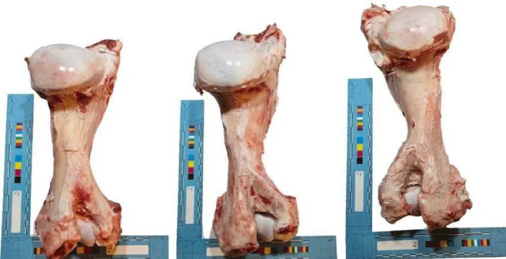
Figure 1: Bones 1, 2, and 3, respectively, before burning.
According to Symes and L’Abbé [5], the most common instrument responsible for sharp-force injuries in forensic cases is a standard kitchen knife. For this reason, a kitchen knife with a pointed tip and double-sloping edges was selected in the present study.
The length, width, direction, and interrelationships of each incision were measured with a forensic scale, photographed, and recorded. Subsequently, each bone was burned separately under open-air conditions using oak wood, without fuel or additives, at 460.0 °C – 570.0 °C for 60 minutes, as measured by an infrared thermometer. The burning procedure was conducted in a standard 90.0 L garden incinerator with the front cover removed. To maintain a steady temperature and flame, a combination of coal, wood, and kindling was used. No accelerant was applied to the bones, as this could have left residues and complicated the analysis [6].
After one hour of burning, the specimens were immediately photographed, and despite advanced combustion and destruction, the incisions that remained macroscopically visible were remeasured, photographed, and recorded. Charred hard tissues were then examined for discoloration and areas of thermal fracture. The post-burning incision lengths were measured again, and the similarities and differences between sharp-force injuries before and after burning were evaluated. The obtained data were compared with the existing literature. Table 1 illustrates how the specimens were numbered.
| Table 1: Numbering of the bones and localization of the incisions. The numbering of the bones used in the study, the number of incisions created in each bone, and their locations on the bone are presented. In the first bone, a single incision was made on the central shaft; in the second bone, two incisions were made on the proximal and distal shafts; and in the third bone, six incisions were created on the central shaft at intervals of 0.5 – 1.0 cm. | |
| Bone No. | Number and location of incisions |
| 1 | One incision on the central shaft |
| 2 | Two incisions on the proximal and distal shafts |
| 3 | Six incisions on the central shaft at 0.5 – 1.0 cm intervals |
Fractures of the bones and the morphological features of the incisions were described. In addition, the pre- and post-burning measurements of incision length and width were compared using the Wilcoxon Signed Rank test. This analysis was performed both for all incisions collectively and for the six incisions created on bone number 3. Statistical analyses were carried out using SPSS version 29.0 (IBM Corp., New York, USA).
The statistical analysis was performed to assess the effect of burning on incision dimensions. Given the limited sample size and the absence of a normal distribution pattern as verified by the Shapiro–Wilk test, non-parametric statistical methods were deemed appropriate for this dataset. Accordingly, the Wilcoxon Signed Rank Test was applied to compare paired pre- and post-burning measurements of incision length and width. The use of non-parametric tests ensured robustness of the statistical evaluation by minimizing the influence of distributional assumptions, thereby providing a more reliable interpretation of the results despite the small sample size.
It was observed that all bones exposed to heat turned gray after 60 minutes and acquired a markedly brittle structure. Despite the advanced degree of thermal damage, all identifiable incisions remained visible to the naked eye. Due to the effect of thermal fractures, loss of periosteal and superficial cortical tissue was noted in the most distal incision created on the shaft of bone number 3. Therefore, the evaluation was conducted on the remaining five incisions. The incisions on the bones appeared more distinct after burning, and a small number of linear fissures radiating from the cuts toward the periphery of the bone tissue were observed (Figure 2). In particular, the margins of the incisions acquired a sharper and drier appearance due to heat, making them more easily distinguishable to the naked eye.
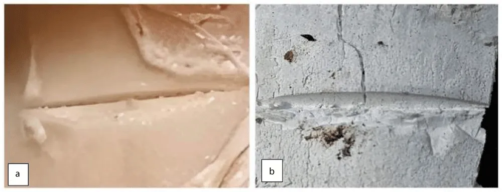
Figure 2: In bone number 1, the periphery of the created incision (a) shows a linear fissure line (b) oriented perpendicularly to the incision, formed after burning.
Table 2 presents the changes in the lengths and widths of the incisions on the bones before and after burning. When all incisions were evaluated together, both the lengths and widths of the bone incisions showed statistically significant increases after burning (p = 0.011 and p = 0.017, respectively). When the same analysis was repeated only for the incisions created on a single bone (bone number 3), the lengths again demonstrated a significant increase (p = 0.042), whereas the increase in widths was not statistically significant (p = 0.066) (Table 2).
| Table 2: Comparison of measurements of artificial incisions created on bones before and after burning. a: Analysis results for all incisions across the three bones. b: Analysis results for incisions on bone number 3 only. W: Wilcoxon test statistic. p < 0.05 was considered statistically significant. | ||||
| Bone No. | Incision Length (Before Burning) (cm) | Incision Length (After Burning) (cm) | Incision Width (Before Burning) (cm) | Incision Width (After Burning) (cm) |
| 1 | 2.00 | 2.10 | 0.15 | 0.21 |
| 2 | 2.10 / 1.50 | 2.30 / 1.85 | 0.10 / 0.08 | 0.15 / 0.10 |
| 3 | 1.00 / 0.70 / 1.60 / 1.30 / 1.20 / 1.00 | 1.30 / 1.00 / 1.70 / 1.50 / 1.45 / — | 0.10 / 0.20 / 0.10 / 0.20 / 0.15 / 0.20 | 0.30 / 0.25 / 0.10 / 0.25 / 0.30 / — |
| Mean ± SD (a) | 1.38 ± 0.47 | 1.65 ± 0.43 | 0.14 ± 0.05 | 0.20 ± 0.08 |
| W (p) (a) | — | 36.000 (p = 0.011) | — | 28.000 (p = 0.017) |
| Mean ± SD (b) | 1.13 ± 0.31 | 1.39 ± 0.26 | 0.16 ± 0.05 | 0.24 ± 0.08 |
| W (p) (b) | — | 15.000 (p = 0.042) | — | 10.000 (p = 0.066) |
In addition to the incisions, longitudinal, transverse, curved transverse, step, and patina fractures were also identified in the burned bones. A marked degree of warping was also observed in the bones, which had become particularly brittle due to heat exposure.
The results demonstrated that sharp-force trauma marks on bone are not completely obscured by heat exposure and that, with further development of such experiments, there is potential for standardization of fracture analysis in burned bones. In the literature, it has been reported that trauma occurring at the time of death may be further exacerbated by heat [7,8]. This has been attributed to the extension and deepening of existing fracture lines originating from impact points along the bone shaft. Indeed, in a 2017 study using 20 sheep radii, Macoveciuc et al. showed that regions traumatized by sharp instruments became more distinct and more clearly observable after thermal alteration, with an increase in incision depth [9].
Typical changes observed in bone following exposure to fire include weight loss, discoloration, and fracturing. Bone generally first turns black (charred), then gray, and finally becomes completely white (calcined) as organic components are consumed [6,10-13]. Thermal fractures can be distinguished from trauma-related fractures by their irregular nature and their tendency to follow the microstructure of the bone [14].
In our study, observations also revealed a small number of linear fissures extending peripherally from the incisions, attributed to the effects of thermal damage on the bone tissue. Thermal damage caused fractures, initiating around the incisions to spread to larger areas, compromising the overall integrity of the bone and intensifying the thermal damage in these regions. Moreover, fractures were also observed in non-incised areas due to high heat exposure. This indicates that the effects of cuts and thermal fractures may occur independently. In particular, in bone number 3, secondary fractures were observed after burning in the areas between the incisions (Figure 3).
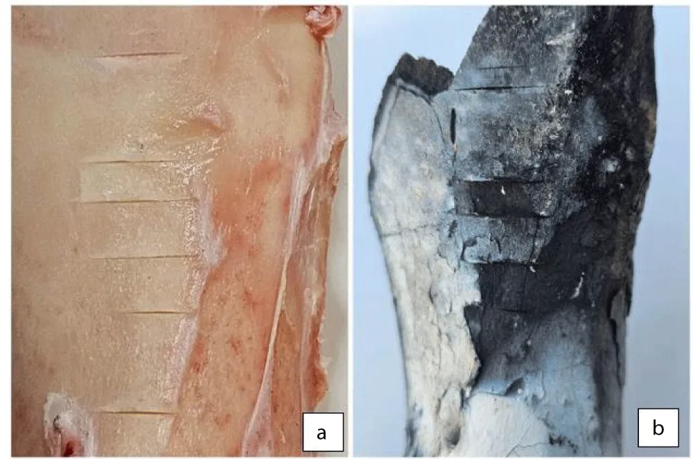
Figure 3: Macroscopic changes observed in Bone 3 before burning (a) and in the areas between the incisions after burning (b).
The observed pattern of thermal damage is thought to be related to the deterioration of bone structure and the rapid loss of water content from the impacted areas, rendering the bone brittle. Indeed, early studies in the literature support our observations, demonstrating that the dehydration process reduces both the strain at fracture and the energy required for fracture in bone [15-17]. Furthermore, as trabecular bone loses water, its buckling behavior—reflecting its capacity to deform under load—shifts from flexible to brittle, making it prone to cracking [18]. The loss of water and elasticity is therefore expected to increase the susceptibility of bone to fracture.
The fractures observed in our study encompassed nearly all types of thermal fractures described by Symes and L’Abbé [5], including longitudinal, transverse, curved transverse, step, and patina fractures. These fractures were also evident in the areas between the incisions in bone number 3, which may explain why no significant difference was observed in incision widths.
The use of bovine rather than human bone and the application of incised wounds rather than other types of trauma represent important limitations of our study. It is known that while the cortical thickness of the human humerus accounts for approximately one-quarter of the total diameter, the cortical thickness of the bovine humerus accounts for nearly half, with a higher density [19]. Moreover, in real-life scenarios, the long bones we studied are more likely to be injured by sharp-blunt instruments rather than sharp-penetrating instruments. Even if they are wounded by sharp-penetrating instruments, the puncture characteristics may predominate in such injuries [20,21]. In addition, in this study, trauma was applied postmortem and without the surrounding soft tissues, including muscle, fat, and skin, covering the bone. Therefore, our study is limited in scope, highlighting the direction and characteristics of the observed changes.
Heat exposure led to a reduction and shrinkage of bone mass, which in turn increased the width and length of the incisions. Therefore, when measuring incisions on burned bones, caution must be exercised to avoid overestimating the blade width of the sharp instrument used. The findings from bone number 3 demonstrated that when multiple sharp-force injuries are present on a single bone, localized thermal damage may also become more severe.
The results of this study indicate that, to some extent, the locations of sharp-force injuries can be recognized by examining the thermal damage patterns on skeletal remains; however, the accurate determination of the blade width and length of the instrument used remains limited. Additional variables should be examined, and further research is needed to assess the post-burning visibility of sharp-force trauma on bone and to enable more reliable identification of the instrument employed.
Author contributions:
Conceptualization: AA
Methodology: AA
Data collection: AA.
Analysis and interpretation: AA.
Literature review: AA.
Writing – original draft: AA.
Writing – review and editing: AA.
Final approval of the manuscript: AA.
- Vakfı U. Türkiye Armed Violence Map 2022 Report. 2022. Available from: https://umut.org.tr
- Isenhour JL, Marx J. Advances in abdominal trauma. Emerg Med Clin North Am. 2007;25(3):713–33. Available from: https://doi.org/10.1016/j.emc.2007.06.002
- Hürriyet. Turkey's violence map. 2016.
- Doğan KH, Demirci Ş, Koç S. Killed first, burned after: attempting to eliminate victims of homicide by burning. 2011;16(2):49–53. Available from: https://www.researchgate.net/publication/281558241
- Symes SA, L'Abbé EN, Chapman EN, Wolff I, Dirkmaat DC. Interpreting traumatic injury to bone in medicolegal investigations. In: Dirkmaat DC, editor. A Companion to Forensic Anthropology. Chichester, West Sussex: Wiley-Blackwell. 2012;340–89. Available from: https://www.researchgate.net/publication/260835132
- Borusiewicz R, Zięba-Palus J, Zadora G. The influence of the type of accelerant, type of burned material, time of burning, and availability of air on the possibility of detection of accelerant traces. Forensic Sci Int. 2006;160(2–3):115–26. Available from: https://doi.org/10.1016/j.forsciint.2005.08.019
- Grévin G, Bailet P, Quatrehomme G, Ollier A. Anatomical reconstruction of fragments of burned human bones: a necessary means for forensic identification. Forensic Sci Int. 1998;96(2–3):129–34. Available from: https://doi.org/10.1016/s0379-0738(98)00115-7
- Collini F, Amadasi A, Mazzucchi A, Porta D, Regazzola VL, Garofalo P, et al. The erratic behavior of lesions in burnt bone. J Forensic Sci. 2015;60(5):1290–4. Available from: https://doi.org/10.1111/1556-4029.12794
- Macoveciuc I, Márquez-Grant N, Horsfall I, Zioupos P. Sharp and blunt force trauma concealment by thermal alteration in homicides: an in-vitro experiment for methodology and protocol development in forensic anthropological analysis of burnt bones. Forensic Sci Int. 2017;275:260–71. Available from: https://doi.org/10.1016/j.forsciint.2017.03.014
- Bradtmiller B, Buikstra JE. Effects of burning on human bone microstructure: a preliminary study. J Forensic Sci. 1984;29(2):535–40. Available from: https://pubmed.ncbi.nlm.nih.gov/6726157/
- Correia PM. Fire modification of bone: a review of the literature. In: Haglund WD, Sorg MH, editors. Forensic taphonomy: the postmortem fate of human remains. 1997;275–93. Available from: https://www.researchgate.net/publication/313078830
- Glassman DM, Crow R. Standardization model for describing the extent of burn injury to human remains. J Forensic Sci. 1996;41(1):152–4. Available from: https://pubmed.ncbi.nlm.nih.gov/8934717/
- Ubelaker DH. The forensic evaluation of burned skeletal remains: a synthesis. Forensic Sci Int. 2009;183(1–3):1–5. Available from: https://doi.org/10.1016/j.forsciint.2008.09.019
- Herrmann NP, Bennett JL. The differentiation of traumatic and heat-related fractures in burned bone. J Forensic Sci. 1999;44(3):461–9. Available from: https://pubmed.ncbi.nlm.nih.gov/10408102/
- Dempster WT, Liddicoat RT. Compact bone is a non-isotropic material. In: Proceedings of the Third U.S. National Congress of Applied Mechanics; 1952 Jun 18–21; Purdue University. New York: ASME. 1952.;123–33. Available from: https://doi.org/10.1002/aja.1000910302
- Evans FG. Mechanical Properties of Bone. Springfield (IL): Charles C. Thomas. 1973;316. Available from: http://www.oandplibrary.org/al/1969_01_037.asp
- Smith J, Walmsley R. Factors affecting the elasticity of bone. J Anat. 1959;93(Pt 4):503. Available from: https://pubmed.ncbi.nlm.nih.gov/13832048/
- Townsend PR, Rose RM, Radin EL. Buckling studies of single human trabeculae. J Biomech. 1975;8(3–4):199–201. Available from: https://doi.org/10.1016/0021-9290(75)90025-1
- Watson JT, McClelland JA. Distinguishing Human from Non-Human Animal Bone: A Guide for Non-Specialists. Tucson (AZ): Arizona State Museum, University of Arizona. 2018;35. Available from: https://statemuseum.arizona.edu/sites/default/files/Distinguishing%20Human%20From%20Animal%20Bone%20%28Watson%20and%20McClelland%202018%29.pdf
- Ekizoğlu O, KS. Yaralar. In: Forensic Medicine in Primary Care [Internet]. Istanbul: Istanbul Medical Chamber. 2011;65–7.
- Aksoy E, ÇA, Ege B, Günaydın G, İnanıcı MA, Karali H, Karagöz M, Ötker C, Yemişçigil A. Forensic Traumatology. 1999. Available from: https://www.ttb.org.tr/eweb/adli/4.html